Home > Research and development > Medical applications
Medical applications
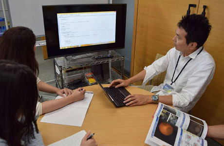
Medical research using composite optical fiberscope systems
Fusion of engineering and medicine has resulted in minimally invasive procedures such as endoscopic surgery or robotic surgery, and these procedures are being performed actively in recent years. The prevalence of minimally invasive procedures leads to lower patient stress and shorter stays in the hospital, contributing greatly to an improvement in the patients' quality of life.
The composite optical fiberscope takes advantage of the composite optical fiber for its ability to simultaneously transmit image data and laser over a single line. It offers significant merit to minimally invasive surgical procedures, whose increased availability continues to be sought after, and also has potential for various other applications.
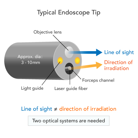
Characteristics of the composite optical fiberscope
Using a typical endoscope requires significant skill and experience, because the direction of observation differs from that of the laser beam.
However, in a composite optical fiber, the direction of observation matches the direction of the laser beam. The operator can then see the area of interest while irradiating the laser more easily and safely. The composite optical fiberscope is also more compact than a typical endoscope, enabling minimally invasive treatment.

Examples applications
Observation of peripheral airways
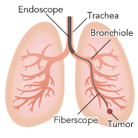
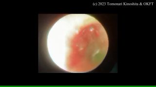
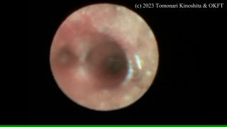
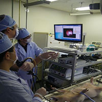
OK Fiber Technology Co., Ltd. has developed a composite optical fiberscope with an outer diameter of 1 mm that can be inserted into the peripheral lung field for observation and irradiation. This fiberscope was able to reach the alveoli, which cannot be reached by conventional bronchoscopes, and observe all airways from the central airways to the alveoli. The following three movies show the actual lumen of a human airway using surgically-resected lungs. We hope to apply this technique to efficient biopsy and treatment of tumors in the future.
The following videos are provided by Dr.Kinoshita, Tachikawa Hospital.
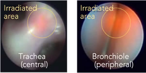
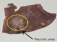
Treatment of peripheral lung cancer
Intrauterine procedures and examination
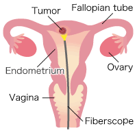
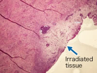
Cervical cancer forms at or near the entrance of the uterus (the cervix), and partial removal of the cervix may enable complete recovery while still maintaining fertility. On the other hand, endometrial cancer, which forms inside the uterus (uterine body), is normally treated through a complete hysterectomy even at early stages, resulting in loss of fertility. The number of new cases of endometrial cancer has continued to increase, exceeding 8,000 in 2005. Looking at the different age groups, the number of cases peaks at the 50-60 year age group, but there is still a significant number around 40 years old, before menopause. With a declining birth rate and an aging population, and with marriage coming later in life, people want treatment options that avoid a complete hysterectomy.
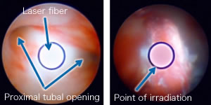
Normally, when examining or treating the uterus, the cervix must be dilated to secure an insertion path for the surgical device. Our composite optical fiberscope with an outer diameter of 1.1 mm (capable of transmitting laser through the fiber core) can be inserted without dilating the cervix, greatly reducing the stress on the patient.
Also, by taking advantage of the fiber's ability to guide multiple laser beams with different wavelengths, we are envisioning a hybrid diagnosis/treatment in which cauterization, photodynamic diagnosis (PDD), and photodynamic therapy (PDT) can be conducted with a single optical fiberscope.
Examination inside the small intestine
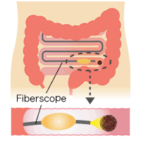
Development of an endoscope for the small intestine that can be used on patients with intestinal obstruction or adhesion
- Enables continuous treatment and diagnosis of the small intestines
- Clinical research is ongoing
Examination inside the pancreas and common bile duct
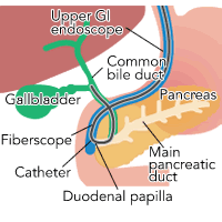
Examination of the inside of the pancreas and common bile duct, as well as treatment of gallstones, pancreatic calculi, and cancers of the gallbladder and pancreas
- Enables observation of narrow spaces where insertion of a cholangioscope is difficult
- Can be inserted into the main pancreatic duct and the common bile duct for internal examination without making an incision in the duodenal papilla
Non-interruptive vascular bypass surgery tool
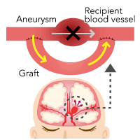
Creating a bypass without interrupting the blood supply within the blood vessels
- Sever the vascular wall under observation
- Currently constructing a non-interruptive vascular bypass system
- 2012 to present: Preclinical tests started
Endoscopic neurosurgery tool
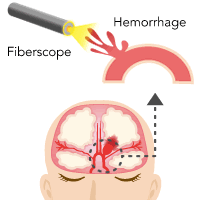
Development of an endoscopic neurosurgery device that can accurately stanch blood
- Enables selective blood stanching
- 2012 to present: Preclinical tests started